
With this extra space and without the discs acting as a cushion, the facet joints cannot control the movement of the vertebrae as well and they become hypermobile which can, eventually, lead the a vertebrae disc moving forward. As the discs lose water, they shrink which brings the vertebrae above and below, along with their facet joints, closer together. Over time, vertebrae discs naturally begin to lose water as their outer wall, known as the annulus, weakens.

TYPE IIIĪging and disc degeneration are the main culprits in type III spondylolisthesis. However, instead of long-term overuse and hyperextension, the break is caused through intense trauma (like a car accident).
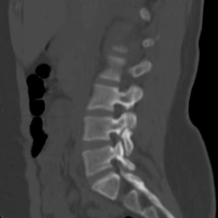
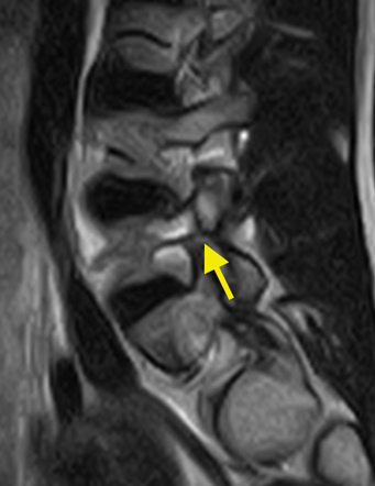
#Pars defect with spondylolisthesis full
Type II C: Like type II A, this condition does involve a full fracture of the pars interarticularis. A longer pars can cause the vertebrae to slip forward. Instead, new bone grows in an attempt to heal the damage which causes it to stretch. Type II B: This form of spondylolisthesis is similar to Type II A, however in this condition the pars interarticularis fractures, but does not fully break. In this condition, the pars fractures completely which allows the injured vertebrae to shift or slip forward. Type II A: This form of Type II spondylolisthesis commonly occurs in people who engage in high intensity, contact sports like football, gymnastics and weightlifting and have multiple microfractures in the pars interarticularis due to overstretching (hyperextension) and overuse.

Type II spondylolisthesis can be divided into three individual subcategories. In Isthmic II spondylolisthesis there’s a problem with the pars interarticularis, a section of bone that sits towards the back of the vertebrae. Type II spondylolisthesis, also known as isthmic, is the most common form of the condition and it usually occurs in adults as a result of abnormal wear on the vertebrae from repetitive stress. This most commonly occurs where the lumbar spine and the sacrum join, near the L5-S1 area. However, in type I spondylolisthesis, the facet joints don’t work properly and allow a vertebra to slip. They are largely responsible for stability and movement within the spine they keep the bones in the spine lined up, while still allowing them to move and remain flexible. Facet joints act much like hinges and run in pairs down the length of the spine, one on each side. Type I spondylolisthesis is often caused by a birth defect in the articular processes of the spine (the parts of the spine that are meant to control movement, like facet joints). Congenital is defined as a disease or or physical abnormality that is present from birth. Type I Spondylolisthesis is also commonly known as congenital spondylolisthesis. Additionally, the muscles in legs may feel tight or weak which can cause patients to walk with a limp. When it does occur, however, it often manifests itself as pain in the lower back or buttocks. Occasionally, a patient can slip their vertebrae and not feel any pain or numbness until years after the fact. However, not all symptoms are immediately prominent. This slippage can lead to pressure on the spinal cord or nerve roots which can cause back pain as well as numbness and weakness in both legs. The most common area for spondylolisthesis to occur is within the bottom level of the lumbar spine between L5-S1. Spondylolisthesis occurs when one of the lumbar vertebrae in the spine moves forward relative to the vertebrae below it, causing pain or weakness. The spinal condition is chronic, meaning it can last for years or be lifelong, but is typically treatable by a neurosurgeon. Spondylolisthesis is a very common cause of back pain in the United States, affecting approximately 3 million Americans every single year.


 0 kommentar(er)
